유방 해부학
유방 (breast)
[유방의 구조]
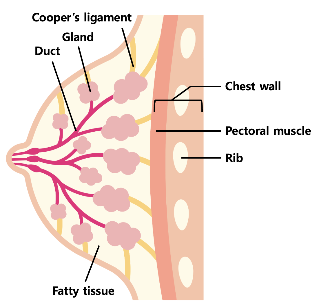
위치: 2nd ~ 6th rib 사이, 흉골(sternum)에서 중앙액와선(midaxillary line)까지 분포
Tail of Spence (Axillary tail): 유방 조직이 액와부(axilla)로 뻗어 나간 부분
Retromammary space: 유방 조직과 대흉근 근막(pectoralis fascia) 사이의 공간 (유방의 가동성 제공)
Cooper’s ligament: 유방을 지지해주는 역할을 하는 인대
임상적 의의: cooper's ligament 주변에 암 등 질병이 생겼을 때 cooper’s ligament가 당겨져서 skin dimpling이 발생할 수 있다.
[혈액 공급 (Blood Supply)]
Internal mammary artery (Internal thoracic a.): 천공지(perforating branches)를 통해 약 60% 공급
Lateral thoracic artery: Axillary artery의 분지로 약 30% 공급
[유방의 미세 구조]
: 아래 사진은 유방의 구조에서 gland 부분을 확대한 것이다.
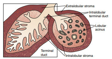
Terminal Duct - Lobular Unit (TDLU)
: 유방에서 유즙을 분비하는 glandular tissue의 terminal duct 와 acinus를 일컫는 용어. 유방암이 발생하는 주요한 구조물
[유방의 초음파 소견]
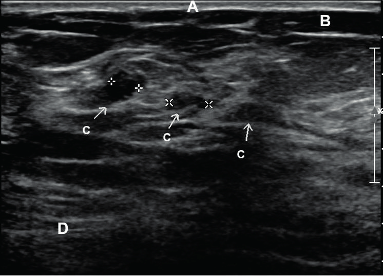
(A) skin, (B) subcutaneous fat, (C) terminal duct lobular unit, (D) muscle
액와 (axilla)
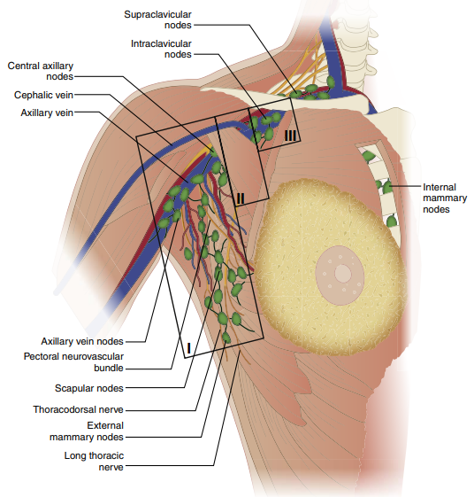
[액와 림프절 (Axillary lymph nodes)]
소흉근(Pectoralis minor m.)을 기준으로 3개의 Level로 분류 (Berg's level)
Level I : lateral border of pectoralis minor m.
Level II : anterior & posterior to pectoralis minor, posterior to pectoralis major
Rotter's node (Interpectoral node): 대흉근과 소흉근 사이에 위치, Level II에 포함됨
Level III : subclavicular/ intraclavicular node
※ 유방 림프의 75% 이상이 액와 림프절로 배액됨 (나머지는 내유 림프절 등으로 배액)
[신경 (nerve)]
Long thoracic nerve: serratus anterior muscle에 innervate하여 scapula를 chest wall에 고정시키는 역할 → 수술 중 손상시 winged scapula(익상견갑, 견갑골이 날개처럼 튀어나와 보이는 것) 발생
연습문제 8문제
0/8 완료
0개의 글
** 제목만 보더라도 어떤 내용인지 알 수 있도록 완성된 문장으로 작성해주세요.
예시) 초음파 (X) → 초음파 사진에서 PDA 소견을 어떻게 알 수 있나요? (O)